
After a recent meeting at a client site, I realised that I was having trouble making out the presentation on the board from my seat in the middle of the room. Having been first diagnosed with myopia as a young child and extremely familiar with an ever-changing prescription, I decided it was time to make an appointment for an eye exam.
I asked my husband if he recommended a particular eye doctor. He corrected me that I needed an “optician” – apparently in the UK the term “eye doctor” isn’t used.
After my minor annoyance of having my English corrected, I decided to google and found a place called Specsavers. Being a fan of online appointment booking, I found a location near me and started the booking. During the process, I was asked if I wanted to have an OCT scan for £10. Friends – I would love to tell you this was a thoughtful decision process, but it was actually “Yeah – why not. It’s only £10.”

So here’s how that went
I do the exam. Really interesting that even across continents, the exam was very similar to the exams I have had over the course of my lifetime. Read the smallest line you can see on the chart – is it better with lens 1 or lens 2?
Sure enough – my prescription had changed. In fact, it changed a LOT – better get those glasses before I try driving across country again. The optician (see me using my fancy new word) started talking to me about different types of lenses, what will be good for being in front of the computer all day, etc. He was so calm and had such a soothing voice.
He then says, let’s look at your OCT scan.
What the heck is an OCT scan?
I hadn’t really heard of an Optical Coherence Tomography (OCT) scan to be honest. According to Specsavers
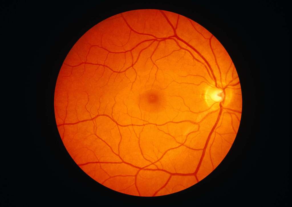
OCT is separate to an eye test. An eye test checks your eye health as well as how well you can see. Part of that often involves taking an image of the back of the eye (digital retinal photography), but an OCT scan takes this a step further, allowing your optician to look even deeper into your eyes and the structures within them.
So he takes a look at my scan and see he is concerned. He moves back and forth between the OCT scan and the digital retinal photography image and tells me he needs to take a closer look at my eye with the microscope. After a thorough examination, he breaks the news to me. He says he suspects my retina has become detached. After a call to Torbay Hospital, it is determined I need to be seen right away – as in that day – for further examination.

The Ophthalmologist Exam
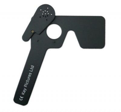
Torbay Hospital has always treated me well – and today was no exception. I learned several new things. One of the (what I considered neato) things is that there is a test called Pinhole visual acuity. What this means is that when my vision was tested (glasses on!) it was drastically improved by the use of a pinhole eye shield, which is super common with myopia.

I underwent another imaging exam. Afterwards, I underwent further examination with a microscope and a special lens placed directly on my eye – this is called Gonioscopy. Finally I was sent to the lead ophthalmologist to provide a diagnosis and review my options.

Asymptomatic Retinal Detachment
The diagnosis is Asymptomatic Retinal Detachment.
The usual causes of retinal detachment are tiny holes or tears in the retina that allow fluid to spread under the retina and lift it away from the back wall of the eye .
It sounds scary because it is. The ophthalmologist shows me the different layers in my eye indicating that the tear has grown over the past few months. He does a demo with my left eye covered to show that my vision is obstructed in the upper left corner of my right eye due to the detachment.
The good news is that it has probably been there a few months. It isn’t a sudden event. The reason this is good (to me, anyway) is because it gives me time to consider my options.
He talks to me about a surgery called Scleral buckle. He explained it in great detail, but I was most interested in the part where he said I would be asleep and not feel anything during it.
I have the option to just monitor it and not do the surgery. The obvious risk is that the tear could grown and as it gets closer to the centre of my eye, it is a bigger threat to my vision.

And there you have it
How does this relate to accessibility? I think it is obvious. Technology and innovation has provided a way for a specialist to see the detail of my eye, including the retina, even though I had no symptoms that eluded to a retinal detachment. I am excited to see how technology becomes more accessible to the ordinary person and helps improve our ability to enjoy the world around us.
